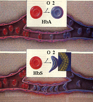
The schematic diagram shows the changes that occur as sickle or normal red cells release oxygen in the microcirculation. The upper panel shows that normal red cells retain their biconcave shape and move through the microcirculation (capillaries) without problem. In contrast, the hemoglobin polymerizes in sickle red cells when they release oxygen, as shown in the lower panel. The polymerization of hemoglobin deforms the red cells. The problem, however, is not simply one of abnormal shape. The membranes of the cells are rigid due in part to repeated episodes of hemoglobin polymerization/depolymerization as the cells pick up and release oxygen in the circulation. These rigid cells fail to move through the microcirculation, blocking local blood flow to a microscopic region. Amplified many times, these episodes produce tissue hypoxia. The result is pain, and often damage to organs.
Recently, red cell adhesion to endothelial cells has been recognized as a major factor in the pathogenesis of sickle cell disease. Normal red cells do not adhere to endothelial cells. In contrast, sickle cells are quite "sticky". Even brief adherence to endothelium would increase the probabilty of the red cells sickling before they can get out of the capillaries into the endothelium. This enhances the chance of vaso-occlusion and local tissue hypoxia.