This page was last modified on:


Hematocrit

Figure 1. Schematic of Hematocrit Determination
Hematocrit Schematic
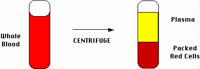
To determine the hematocrit, whole blood in a tube is centrifuged to pellet the red cells (packed red cells). Plasma remains on top of the packed red cells. The fraction of the blood that is packed red cells is the hematocrit. In this example, the hematocrit is about 40%.
Blood has a liquid component (plasma) and a particulate component (blood cells). The denser blood cells (most of which are red blood cells) will settle in a tube, particularly if the blood is spun in a device called a centrifuge. The fraction of the column composed of red blood cells relative to the entire column is the hematocrit, which normally is in the range of 40%. The upper portion is yellowish plasma. Red blood cells carry oxygen. Blood with a low hematocrit (e.g., 20%) leaves the tissues relatively oxygen-starved and weak. A high hematocrit (e.g., 70%) produces problems as well, but is uncommon.


Bibliography

Laurence Corash, "Laboratory Hematology: Methods for the Analysis of Blood", in Blood: Principles and Practice of Hematology, eds.

Robert I. Handin, Samuel E. Lux, Thomas P. Stossel (Philadelphia: J.B. Lippincott Company, 1995) 23