revised October 8, 1999

Chelators for Iron Overload

What are Chelators?

 Chelators are small molecules that bind very tightly to metal ions. Some chelators are simple molecules that are easily manufactured (e.g., ethylene diamine tetra acetic acid; EDTA). Others are complex proteins made by living organisms (e.g., transferrin). The key property shared by all chelators is that the metal ion bound to the chelator is chemically inert. Consequently, one of the important roles of chelators is to detoxify metal ions and prevent poisoning. For instance, EDTA is used to treat patients with extreme, life-threatening hypercalcemia. The iron chelator, desferrioxamine, is used to remove excess iron that accumulates with chronic blood transfusions.

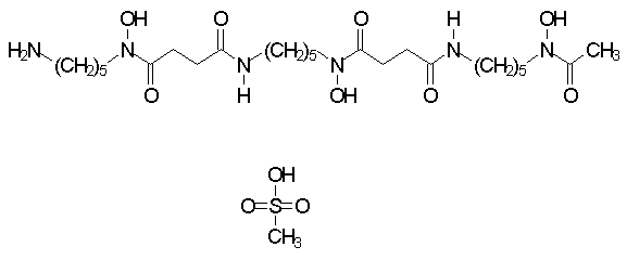
 Many chelators are used in chemistry and industry. Only a few are clinically useful since most have dangerous side-effects. One important property required of clinically useful chelators is specificity. Since these drugs disperse diffusely in the body, they must bind the target metal ion preferentially over others. Desferrioxamine (Desferal®), for instance, can be used to treat iron overload since the drug binds iron with a large preference over other metal ions such as calcium (Kd=10-31 M for iron, Kd=10-9 M for calcium) (1).

Iron Chelators

 Iron chelators can be classified using a number of criteria such as their origin (synthetic versus biologically produced molecules), their interaction with solvents such as water (hydrophobic versus hydrophilic) or their stoichiometric interaction (bidentate versus hexadentate.) Some of these properties have an important impact on the clinical utility of a chelator, as discussed later.

 One key clinical feature of iron chelators is the degree to which they are absorbed from the gastrointestinal tract. A clinically highly effective iron chelator such as desferrioxamine has the drawback of very poor absorption from the gastrointestinal tract (2). Consequently the drug must be given parenterally, as a continuous subcutaneous infusion, or as a continuous intravenous infusion (3, 4). The expensive medical paraphernalia required for desferrioxamine administration makes the treatment expensive, and curbs its availability in areas of the world where medical resources are limited. Even when the resources exist to support iron chelation with desferrioxamine, the intrusiveness of pumps and other paraphernalia often impedes patient compliance (5). For these reasons, an intensive search for orally active iron chelators is being conducted by a number of medical researchers.

Iron Toxicity

 The goal of iron chelation therapy is to prevent iron-mediated injury to cells. The basis of this injury is the very property that makes iron vital to all life: it can exist in either of two stable oxidation states. Iron ions in aqueous solution exist either in the ferrous (Fe2+) state or the ferric (Fe3+) state. The shift of electrons between iron and donor molecules is the basis of energy production by controlled oxidation of carbohydrates, proteins, and lipids. Iron is a key element in most of the cytochrome enzymes involved in the oxidative phosphorylation of the Krebs cycle.

 Because of its ability to participate in chemical reactions that involve the shift of electrons between molecules (reduction-oxidation or redox reactions), the body tightly regulates iron. When iron is tightly bound to a chelator molecule, be it a protein or a small chemical, the reactivity of the iron is greatly dampened. The key iron storage protein in the body is ferritin. Ferritin is a very large spherical molecule (6). Iron is deposited as semi-crystalline deposits inside these protein "vaults". Iron that is sequestered within ferritin is metabolically inactive.

 The iron deposits in patients who have received multiple blood transfusions for chronic anemia, such as thalassemia, can exceed the storage and detoxification capacity of ferritin. Consequently, "free" (or more accurately, loosely bound) iron begins to accumulate in tissues and blood. This "free" iron can catalyze the formation of very injurious compounds, such as the hydroxyl radical (.OH) from compounds such as hydrogen peroxide, which are normal metabolic byproducts (Fenton reaction) (7).

 The hydroxyl radical is highly reactive, and attacks lipids, proteins and DNA (8). The initial reaction with each of these molecules is the formation of peroxides (e.g., lipid peroxides) that can interact with other molecules to form cross links. These cross-linked molecules perform their normal functions either poorly or not at all.

Lipids
 Peroxidaiton promotes cross links in membrane lipids, creating islands or domains of dysfunctional molecules. Cell membranes, which consist primarily of lipids, stiffen and acquire odd shapes. This is particularly problematic for red cells, which have no nucleus. Unlike most other cells, red cells cannot repair membrane damage. The red cells of patients with thalassemia or sickle cell disease loose the elasticity needed to pass through the microcirculation (9). For patients with sickle cell disease, this exacerbates the problem of microvascular vaso-occlusion. These damaged red cells are removed by reticuloendothelial cells, most prominently in the spleen.

Proteins
 Protein cross linking can create protein clusters, particularly in membranes (10,11). Again, red cells are particularly susceptible to such damage, lacking membrane repair mechanisms. The cells of the immune system recognize these protein clusters as being abnormal. Antibodies to these clusters (termed "membrane senescence antibodies") promote removal of damaged red cells from the circulation. The result is enhanced hemolysis. Oxidation of band 3, the red cell anion transport channel, disturbances the osmotic balance of red cells and impairs their function.

DNA
 DNA cross-links can impair cell replication, leading to cell death. The degree of cross-linking produced by reactive oxygen species in patients with iron overload generally is relatively small and probably relatively unimportant.

 Although red cells are very susceptible to iron-mediated cell injury, they do not bear the assault of reactive oxygen species alone. Damage to cells in other organs accumulates gradually, and eventually becomes clinically significant. Hepatocytes, the primary component cells of the liver, are the major storage site for body iron. With iron overload, these cells are relentlessly bombarded by reactive oxygen species and eventually die (12). They are replaced by fibroblast cells. The collagen laid down by fibroblasts produces liver fibrosis and, eventually, cirrhosis.

 Likewise, cardiac cells are damaged with iron overload (13). Normal cardiac function requires the coordinate activity of all the cells in the heart. Damaged, poorly-functioning cells often fail in this regard. The clinical manifestations include congestive heart failure (due to injury to myocytes) and arrythmias (due to damage to the cells of the cardiac conducting system) (14, 15). Either can be deadly.

Protection of Cells by Iron Chelators

Chelators protect cells from iron-mediated toxicity in two ways.  The most readily apparent mechanism by which chelators provide protection is removal of the excess iron from the body. Once the toxic iron is gone, the body's repair mechanisms can swing into action to correct damage that may have occurred. The ability of chelators to remove excess iron depends on (at least) two factors: (a) the rate at which the chelator depletes storage iron, and (b) the rate of continued iron accumulation.

 Patients with some disorders develop iron overload due to repeated transfusions, afterwhich the underlying disorder is corrected. For instance, some people with aplastic anemia who receive a bone marrow transplant require many transfusions for support until the graft matures. Thereafter, they have a normal hematocrit. Chelators can remove all the excess iron in this setting.

 Most patients with transfusional iron overload require transfusions indefinately. Examples of this include people with thalassemia major and some froms of myelodysplasia. Since each unit of blood deposits about 230 mg of iron, most patients who require, for instance, 2 units of blood per month will have at most a very slightly negative iron balance with chelation therapy. The most widely used iron chelator, desferrioxamine, removes somewhere between 30 and 70 mg of iron per day. Protection of patients with ongoing transfusion requirements solely by removal of excess iron is uncommon.

The tight binding of chelators to iron blocks the ion's ability to catalyze redox reactions. Iron ions have six electrochemical coordination sites. Consequently, a chelator molecule that binds to all six sites completely inactivates the "free" iron. Such chelators are termed "hexidentate", of which desferrioxamine is an example. With some chelators, a single molecule interactions with only two of the coordination sites on iron. These chelators are called, "bidentate". An example of this type of molecule is ferrichrome. Three molecules coordinate with a single iron ion to produce complete chemical immobilzation. Another example is deferiprone, or "L1", a chelator currently in clinical trials.

 Since neutralization of free iron is essential to protect cells, a molecule such as desferrioxamine has the advantage of inactivating iron as part of a 1:1 molecular complex. On the other hand, bidentate chelators (C) can produce partial reaction products with iron (Fe):

  1. Fe(C) [redox reactive]
  2. FeC2 [redox reactive]
  3. FeC3 [inactive]

With a bidentate iron chelator, a spectrum of chemical species will exist, of which a minority is inactive. A large chemical excess of chelator is needed to push the reaction toward completion, the formation of the FeC3 (inactive) product.

Clinical Efficacy of Iron Chelators

 No ideal chelator exists to treat patients with transfusional iron overload. The characteristics of such a compound can be extrapolated from the clinical requirements:
  1. Oral administration.
  2. Good tissue penetration
  3. Easy mobilization of the iron-chelator complex
  4. Inexpensive
  5. Non-toxic
  6. Hexidentate binding of iron ions

Desferrioxamine mesylate
Structure of desferrioxamine mesylate >
 The most widely used chelator currently is desferrixoxamine (16). As shown in the figure, the drug has multiple carbonyl and hydroxyl groups that provide electrons to cooridnate with those in Fe3+. Desferrioxamine chelates iron in a one-to-one ratio. This drug must be given parenterally (either subcutaneously or intravenously), which limits its utility. Desferrioxamine is expensive and requires sophisticated medical devices for administration. Nonetheless, a plethora of data show that scrupulous use of the medication not only prevents progression of iron-induced injury, but also can reverse organ dysfunction (17, 18). Desferal opens the possibilty of long survival for transfusion-dependent pateints, such as thoseÝ with thalassemia.

 Intensive exploration for an orally active iron chelator is currently under way. The only drug actively considered at present is deferiprone, or "L1". Investigators in Canada, the US, Italy, Greece, and India have studied this agent (19, 20, 21). Presently, no consensus exists on the utility of L1. The drug does not mobilize iron as efficiently as desferrioxamine. This shortcoming could, however, be balanced by better compliance. Some serious side-effects, such as agranulocytosis, occur with L1 (22). The degree to which these will limit its utility is presently unclear. Finally, in contrast to the hexidentate chelating capacity of desferrioxamine, L1 is a bidentate chelator. Intermediate chelation products could continue to produce cell and organ injury in patients treated with this drug. The issue of progression of liver damage in patients who use deferiprone is at the center of the current controversy over the drug (23). Further investigation is warranted to determine where L1 fits in the clinical armamentarium.


References

  1. Keberle, H. 1964. The biochemistry of desferrioxamine and its relation to iron metabolism. Ann NY Acad Sci 119:758-775.
  2. Callender ST, Weatherall DJ. 1980. Iron chelation with oral desferrioxamine. Lancet ii: 689-691.
  3. Propper RD, Cooper B, Rufo RR, Nienhuis AW, Anderson W, Bunn HF, Rosenthal A, Nathan, DG. 1977. Continuous subcutaneous administration of deferoxamine in patients with iron overload. N Engl J Med 297: 418-423.
  4. Cohen AR, Mizanin J, Schwartz E. 1989. Rapid removal of excessive iron with daily, high-dose intravenous chelation therapy. J Pediatr 115: 151-155.
  5. Berati S. 1989. Noncompliance with iron chelation therapy in patients with beta thalassaemia. J Psychosom Res 33: 739-745.
  6. Otsuka S, Maruyama H, Listowsky I. 1981. Structure, assembly, conformation, and immunological properties of the two subunit classes of ferritin. Biochemistry 20: 5226-5232.
  7. Gutteridge JMC., Rowley DA, Halliwell B. 1981. Superoxide-dependent formation of hydroxyl radicals in the presence of iron salts. Biochem J 199: 263 - 265.
  8. Bacon BR, Britton RS. 1990. The pathology of hepatic iron overload: a free radical--mediated process? Hepatology 11: 127-137.
  9. Clark MR, Mohandas N, Shohet SB. 1980. Deformability of oxygenated irreversibly sickled cells. J Clin Invest 65: 189-196.
  10. Rank BH, Carlsson J, Hebbel RP.Ý 1985. Abnormal redox status of membrane-protein thiols in sickleÝ erythrocytes. J Clin Invest 75: 1531-1537.
  11. Corbett JD, Golan DE. 1993. Band 3 and glycophorin are progressively aggregated in density- fractionated sickle and normal red blood cells. Evidence from rotational and lateral mobility studies. J Clin Invest 91: 208-217.
  12. Bonkovsky HL. 1991. Iron and the liver. Am J Med Sci 301: 32-43.
  13. Buja LM, Roberts WC. 1971. Iron and the heart. Am J Med 51: 209-221.
  14. Liu P, Olivieri N. 1994. Iron overload cardiomyopathies: new insights into an old disease. Cardiovasc Drugs Ther 8: 101-110.
  15. Schellhammer PF, Engle MA, Hagstrom JW. 1967. Histochemical studies of the myocardium and conduction system in acquired iron-storage disease. Circulation 35: 631-637.
  16. Ley TJ, Griffith P, Nienhuis AW. 1982. Transfusion haemosiderosis and chelation therapy. Clin Haematol 11: 437-445.
  17. Flynn DM, Hoffbrand AV, Politis, D. 1982. Subcutaneous desferrioxamine: the effect of three years' treatment on liver, iron, serum ferritin, and comments on echocardiography. Birth Defects 18: 347-353.
  18. Rahko PS, Salerni R, Uretsky BF. 1986. Successful reversal by chelation therapy of congestive cardiomyopathy due to iron overload. J Am Coll Cardiol 8: 436-440.
  19. Kontoghiorghes GJ, Aldouri MA, Hoffbrand AV, Barr J, Wonke B, Kourouclaris T, Sheppard L. 1987. Effective chelation of iron in b thalassemia with the oral chelator 1,2-dimethyl-3-hydroxypyrid-4-one. Br Med J 295: 1509-1512.
  20. Olivieri NF, Koren G, Hermann C, Bentur Y, Chung D, Klein J, St Louis P, Freedman MH, McClelland RA, Templeton, DM. 1990. Comparison of oral iron chelator L1 and desferrioxamine in iron-loaded patients. Lancet 336: 8726.
  21. Agarwal MB, Gupte SS, Viswanathan C, Vasandani D, Ramanathan J, Desai N, Puniyani RR, Chhablani, AT. 1992. Long-term assessment of efficacy and safety of L1, an oral iron chelator, in transfusion dependent thalassaemia: Indian trial. Br J Haematol 82: 460-466.
  22. al-Refaie FN, Wonke B, Hoffbrand, AV. 1994. Deferiprone-associated myelotoxicity. Eur J Haematol 53: 298-301.
  23. Olivieri NF, Brittenham GM, McLaren CE, Templeton DM, Cameron RG, McClelland RA, Burt AD, Fleming KA. 1998. Long-term safety and effectiveness of iron-chelation therapy with deferiprone for thalassemia major. N Engl J Med 339:417-423.


HomeBWHMGHHMS